In life sciences, the ability to measure the distribution of biomolecules inside a cell in situ is an important investigative goal. Among a variety of techniques, scientists have used magnetic imaging (MI) based on the nitrogen vacancy center (NV) in diamonds as a powerful tool in biomolecular research. However, nanoscale imaging of intracellular proteins has remained a challenge thus far. In a recent study now published in Science Advances, Pengfei Wang and colleagues at the interdisciplinary departments of physics, biomacromolecules, quantum information and life sciences in China, used ferritin proteins to demonstrate the MI realization of endogenous proteins in a single cell, using the nitrogen-vacancy (NV) center as the sensor. They imaged intracellular ferritins and ferritin-containing organelles using MI and correlative electron microscopy to pave the way for nanoscale magnetic imaging (MI) of intracellular proteins.
Increasing existing spatial resolution of biomedical imaging is required to achieve ongoing demands in medical imaging, and therefore, among a variety of techniques, magnetic imaging is of broad interest at present. Magnetic resonance imaging (MRI) is widely used to quantify the distribution of nuclear spins but conventional MRI can only reach a resolution of 1 µm in nuclear spin imaging where the resolution is limited by electrical detection sensitivity. Scientists have developed a series of techniques to break this resolution barrier, including a superconducting quantum interference device and magnetic resonance force microscopy. Nevertheless, these reports require a cryogenic environment and high vacuum for imaging, limiting the experimental implementation and its translation to clinical practice.
A recently developed quantum sensing method based on the nitrogen vacancy center in diamond has radically pushed the boundary of MI techniques at the nanoscale to detect organic molecules and proteins in the lab. Scientists have combined quantum sensing with NV centers and scanning probe microscopy to demonstrate nanoscale MRI for single electron spin and small nuclear spin ensemble while using the NV center as a biocompatible magnetometer to noninvasively image ferromagnetic particles within cells at the subcellular scale (0.4 µm). For example, depolarization of the NV center can be used as a wideband magnetometer to detect and measure fluctuating noise from metal ions and nuclear spins. However, such imaging of single proteins via MI at the nanoscale has not been reported in the single cell thus far.
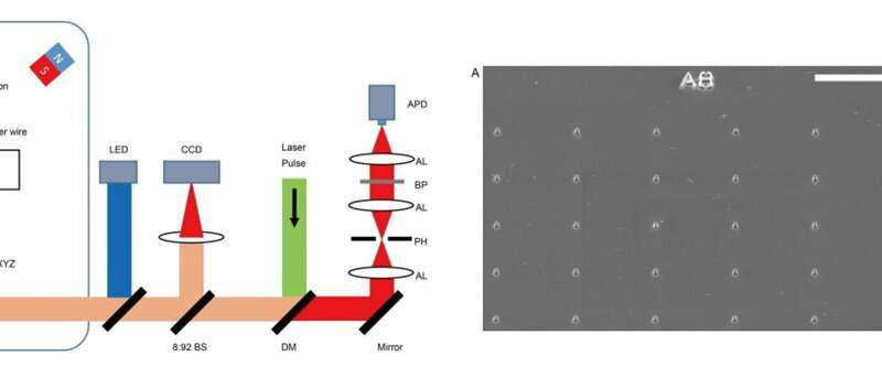
In the present work, Wang et al. reported two technical advancements to allow nanoscale MI of intracellular proteins within a single cell. For this, they freeze-fixed the cell to a solid state and intricately segmented it to a cube shape, then placed it on a tuning fork scanning probe of an atomic force microscope (AFM) for imaging, where the flat cross section of the cell was exposed to air. The scientists used the sample placement setup to allow the NV sensor to be positioned within 10 nm of the target proteins and used the AFM to suppress thermal drift during sample positioning. They then engineered trapezoidal cylinder-shaped nanopillars at a bulk diamond surface for image acquisition, technically shortening the time of image acquisition by one order compared to previous methods. In the present study, the scientists used this technique to conduct in situ MI of the magnetic fluctuating noise of intracellular ferritin proteins (a biomarker of iron stores and transferrin saturation in the body) within the experimental setup.
Ferritin is a globular protein complex with an outer diameter of 12 nm, containing a cavity spanning 8 nm in diameter that allows up to 4500 iron atoms to be stored within the protein. The magnetic noise of the ferric ions can be detected due to their effects on the T1 relaxation time of an NV center. In this work, Wang et al. confirmed the observation using fluorescence measurements of time-dependent decay of the population of NV centers (magnetic spin, mS = 0 state), in a diamond surface coated with ferritins. Additionally, the scientists detected the magnetic noise with label-free methods using the NV center via transmission electron microscopy (TEM). The work allowed the development of a correlated MI and TEM scheme to obtain and verify the first nanoscale MI of a protein in situ.
The scientists used the hepatic carcinoma cell line (HepG2) for the experiments and studied iron metabolism by treating the cells with ferric ammonium citrate (FAC), which significantly increased the amount of intracellular ferritin. They verified this using confocal microscopy (CFM), western blotting and TEM techniques at first. The results showed the primary localization of ferritins in the intracellular puncta around the nucleus, among the cytoplasm. The scientists used bulk electron paramagnetic resonance (EPR) spectroscopy to confirm the paramagnetic properties of ferritin in the FAC-treated HepG2 cells and mass spectroscopy to measure the interference due to other paramagnetic metal ions.
Wang et al. then used ultrafast, high-pressure freezing to immobilize all intracellular components of the Fe-loaded cells. The process stabilized the intracellular structures and molecules by minimizing Brownian motion in cells, which typically contributes to random motion of proteins up to 100 nm in vivo. To image the samples, they embedded and polymerized the frozen cells in LR White medium, followed by gluing the embedded cell sample to the AFM tuning fork with a few cells at the tip. Using a diamond knife, the scientists then sectioned the tip surface to nanometer flatness to examine the cuboid cell section under AFM. They acquired MI images of ferritins by scanning the cell cube along the diamond nanopillars and simultaneously measured NV spin repolarization rate using the “leapfrog” scanning mode of the microscope as detailed previously.
The scientists measured fluorescence decay at a fixed free evolution time of 50 microseconds (τ = 50 μs) to reveal the degree of NV sensor spin polarization, which correlated with the amount of ferritin in the sensing volume. They observed the appearance of some clusters via both TEM and MI images, although some details were not observed in MI, the results confirmed that spin noise from intracellular ferritin contributed to depolarize the NV center. In order to obtain details of the ferritin clusters at higher resolution, the scientists minimized the pixel size to 8.3 nm and acquired MI of high resolution of the proteins as expected.
In this way, Wang et al. explored the sensitivity of NV centers as an appropriate sensor for biological imaging applications at the level of the single molecule. They used the technique as a sensor in the experimental setup to obtain the first MI of a protein at a resolution of 10 nm in situ. The scientists aim to improve the stability and sensitivity of the technique to speed up the scanning process and image a larger area of interest in the cell and locate ferritin beyond the nucleus in association with additional organelles.
The work will contribute to clinical diagnostics to determine biomarker-based iron storage and release in cells. This will include studies on the regulatory mechanisms of iron metabolism during the progression of hemochromatosis, anemia, liver cirrhosis and Alzheimer’s disease. Wang et al. propose to extend the approach in situ to other cellular components with paramagnetic signals, including magnetic molecules, metalloproteins and special spin-labelled proteins. The scientists envision that further studies will explore additional targets suitable for high-resolution MI and correlated TEM imaging techniques, with optical microscopy detection incorporated to the experimental setup to extend the work and determine protein nuclear spin MRI as well as perform three-dimensional cell tomography.