Laser-scanning microscopes can be miniaturized to image microenvironments in vivo via inclusion inside optical micromechanical system (MEMS) devices to replace the existing larger components. Multifunctional active optical devices are emerging components that support miniaturization for diffraction-limited performance with simpler optical system designs in optical devices. In a recent study, Tianbo Liu and a team of researchers in the departments of Electrical and Computer Engineering and Dermatology in the U.S. proposed a catadioptric (allowing both light reflection and refraction) microscope objective lens, featuring an integrated MEMS device to perform biaxial scanning, axial focus adjustment and control spherical aberration.
The materials scientists included a reflective MEMS scanner into the MEMS-in-the-lens architecture to support high-numerical-aperture (NA) imaging that gathers light across a wider range of angles to generate images. Liu et al. implemented the MEMS-in-the-lens architecture by including the scan mirror into the objective lens, where the beam axis was normal to the mirror surface without the need for a beam splitter to separate the incident and reflected beam. They demonstrated the optical performance of the catadioptric system (an optical system that allows both light refraction and reflection with minimal aberration) by imaging hard and soft targets using a confocal microscope based on the new objective lens design. The improved imaging technique will allow advanced diagnosis of medical conditions. The results of the study are now published on Light: Science & Applications.
Unprepared and uncleared organs in live animals can be imaged in vivo using scanning laser confocal and multiphoton microscopy techniques. Technical advances have facilitated bench top imaging of small animal models such as mice, with suitable medical applications also emerging in dermatology clinics to noninvasively examine optical skin biopsies. However, conventional laser scanning microscopes are large and limit both medical and live animal imaging procedures. To access the human body and image ambulatory animals, scientists must therefore miniaturize these instruments.
Miniaturized scanning mechanisms with smaller instruments such as micromechanical system devices can replace existing bulky mechanisms required to scan and focus the beam for hitherto improbable applications. For example, scientists were able to mount a MEMS-scanned miniaturized two-photon microscope weighing only 2.15 g on the head of a freely moving mouse for brain imaging. The devices have also facilitated laser scanning microscopy to be adapted in endoscopic platforms and during MEMS-based optical biopsy experiments to detect cancer in vivo. Alongside its smaller footprint, a MEMS scanner contributes to miniaturization by combining multiple degrees of freedom during its production alongside its optical architecture.
In the present work, Liu et al. explored a new optical architecture for a miniature, high-NA scanning laser microscope with a 3-D MEMS scanner within the objective lens. They illustrated the optical layout of the MEMS-in-the-lens to fabricate the device and operate it in vivo. The scientists engineered the MEMS 3-D scan mirror by successfully reproducing a method previously introduced by the same group. For in vivo microscopy, they operated the hyperhemisphere (that offers a broader field of view) in contact with tissue containing a variable index of refraction ranging from 1.3 to 1.4. Based on the parameters, the scientists simulated the imaging performance of the setup. They concluded hyperhemisphere of BK-7 glass to be effective as a front lens element for a tissue microscope with an active 3-D MEMS scanner deployed at the simulated aperture.
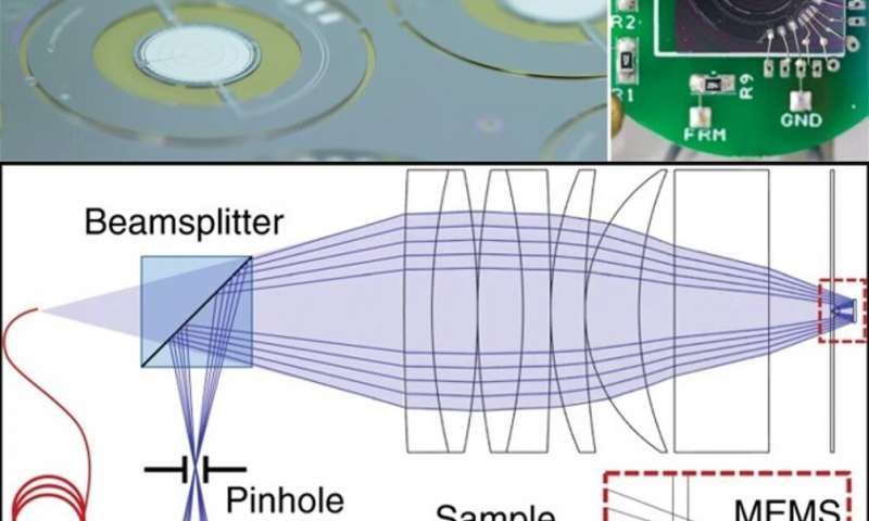
To demonstrate confocal imaging, the scientists used a bench-top mockup of the objective lens with an integrated 3-D MEMS mirror. Liu et al. attached the mirror onto the sample stage using a thin-layer of water-based ultrasound gel. As an example, they introduced samples of human cheek cells (~ 80 µm) on to the sample stage, and captured their images using the microscope thereafter. During imaging, the scientists used a 633 nm helium neon laser for illumination. They then attached the sample of interest on the glass wafer opposite the hyperhemisphere lens. Liu et al. included a 50/50 beam splitter between the optical fiber and compound lens element to separate the reflected light, and a 10 µm pinhole to spatially filter the reflected light.
The MEMS confocal microscope also allowed imaging beneath the surface of the sample and Liu et al. demonstrated this via imaging a sample of interest. For the sample, they suspended 6 µm polystyrene microbeads in an ultrasound transmission gel then followed up the imaging process with volumetric reconstruction of the images to better illustrate confocal sectioning at different focal planes. Although the images were well resolved, the scientists observed that the 3-D profiles of the beads were neither uniform nor symmetric requiring further optimization of the technique.
The 3-D MEMS mirror developed provided complete scanning and focus control for the instrument, alongside electronic control of the spherical aberration. The new work showed improved resolution compared with previously described 3-D MEMS mirrors, to allow its inclusion in a compact MEMS-in-the-lens system.
In this way, Tianbo Liu and co-workers proposed and developed a catadioptric MEMS-in-the-lens microscope objective lens and integrated a MEMS 3-D scanner to perform biaxial scanning with controlled spherical aberration during imaging applications. Liu et al. simulated the development of the proposed instrument architecture to indicate considerable promise for future, miniaturized and high-NA laser scanning microscopes for in vivo imaging applications.